

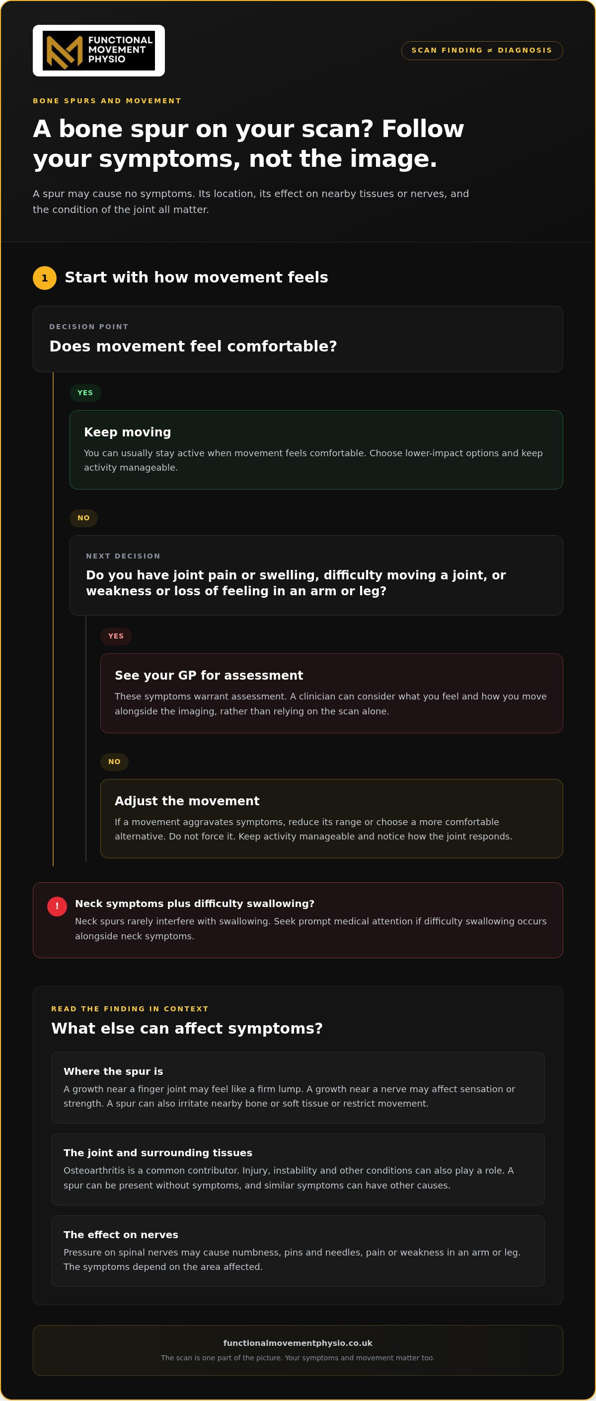
A bone spur on a scan is not, by itself, a reason to stop moving. These bony growths around joints or the spine often cause no symptoms. If movement feels comfortable, you can usually stay active, choosing lower-impact options and adapting movements that trigger pain.
It is understandable to worry that exercise could make a joint worse, especially when pain and stiffness already limit what you can do. But a scan finding does not explain every symptom. Its location, effects on nearby tissues or nerves, and the condition of the joint all matter.
This article explains what bone spurs are, why they develop and how they may affect pain and movement. You will also learn when symptoms warrant medical attention and how to approach activity without pushing through pain. Understanding what may be contributing to your symptoms can help you make informed choices and work towards meaningful movement.
Key Takeaways
- A growth seen on an X-ray may not be the cause of your pain; its location and effect on nearby tissues matter.
- Joint wear and osteoarthritis are common contributors, but other conditions and previous injuries can also play a role.
- Pain, stiffness or changes in movement can have several causes, so focus on understanding the source rather than the scan alone.
- You can adapt activity to your symptoms instead of avoiding movement altogether, choosing options that don’t aggravate pain.
- Support can help you assess movement and build a sustainable path back to the activities that matter to you.
What problems can they cause?
A bone spur may cause no symptoms. The NHS notes that osteophytes can be found on an X-ray taken for another reason. If symptoms appear, their location can influence what you notice. A growth near a finger joint might feel like a firm lump, while one near a nerve may affect sensation or strength. An image alone cannot show whether the growth explains your symptoms.
Pain and stiffness can make everyday movement harder. You may struggle to bend your knee fully, turn your neck comfortably or reach overhead with your shoulder. A spur can irritate nearby bone or soft tissue, restrict movement or press on a nerve. Lumps may also form around the fingers, big toe, heel or ankle.
Pressure on spinal nerves can cause symptoms beyond the joint itself. Depending on the affected area, you may experience numbness, pins and needles, pain or weakness in an arm or leg. Spinal growths can narrow the space around nerve roots or the spinal cord. Neck spurs rarely interfere with swallowing, so seek prompt medical attention if difficulty swallowing occurs alongside neck symptoms.
Symptoms vary by location. Knee osteophytes may accompany difficulty bending or straightening the leg. A hip spur may make movement painful or reduce the joint’s range. A neck spur can affect nearby nerves. Similar symptoms can have other causes, so do not assume an X-ray finding explains every ache or change in movement.
To manage osteophytes, choose lower-impact movement, such as a comfortable walk, and adapt activities that trigger pain. If a movement aggravates symptoms, reduce its range or choose a more comfortable alternative instead of forcing it. Keep activity manageable and notice how the joint responds.
In the UK, see your GP if you have joint pain or swelling, difficulty moving a joint, or weakness or loss of feeling in an arm or leg. These symptoms warrant assessment because joint or nerve problems can affect movement and function. A clinician can consider your symptoms alongside imaging, rather than relying on the scan alone.
According to the NHS, treatment is needed only when a spur causes problems such as pain or stiffness. Nonsurgical care may include physiotherapy to address movement and strengthen supporting muscles. For neck symptoms, treatment may also focus on reducing inflammation and managing pain. The aim is to improve function and address contributing factors, not simply focus on the bony growth.
What causes bone spurs?
Bone spurs develop as bone responds to damage, repeated stress or changes around a joint. Osteoarthritis is the most common cause, but injury, instability and other conditions can also contribute.
Causes and Associated Conditions
A bone spur is a bony growth linked to changes in bone, joints or tendon attachments. Its presence may point to an underlying issue, but does not explain the whole picture by itself. For example, a spur near the knee may accompany joint damage, while one in the neck may relate to structural changes or mechanical stress.
Ageing, degeneration and mechanical instability are associated with spur formation. Previous injury can also prompt the body to form new bone. In the neck, posture problems, alignment changes and reduced structural support may contribute. These factors can interact, so understanding the affected area helps put an imaging finding into context.
Bone spurs may be associated with underlying conditions. Other associations include degenerative disc disease, diffuse idiopathic skeletal hyperostosis, tendonitis and spinal stenosis. These conditions affect different tissues, so an association does not mean that each one causes every spur. The underlying joint or spinal problem matters.
Links to Arthritis
Arthritis, particularly osteoarthritis, is strongly linked to bone spur development. In osteoarthritis, cartilage that cushions the ends of bones breaks down. As the joint changes, the body may form extra bone near the damaged area. This is one reason spurs commonly appear alongside arthritic changes.
As cartilage wears away, the joint’s surfaces become less protected, and movement may place stress on surrounding structures. The body’s response can include new bone and fibrocartilage, a supportive tissue with properties between cartilage and bone. Over time, this process may alter the shape of a joint, such as at the knee or fingers.
Having arthritis raises the likelihood of developing spurs, but the two findings are not interchangeable. A spur can be present without symptoms, while arthritis may affect movement through cartilage loss and other joint changes. Understanding the cause helps explain why treatment needs to address joint function and the underlying condition, not just the bony growth.
How Bone Spurs Form
Bone formation can occur as the body responds to mechanical damage, stress or instability. Around a damaged joint, cells involved in bone growth may build up extra tissue. This can increase the joint’s surface area and change its outline. A spur is a physical response to local conditions, not simply a growth without context.
A bony projection at a tendon or ligament attachment is called an enthesophyte. It differs in location from growths around a joint, though both involve new bone. Repeated loading or tissue damage near an attachment may contribute. For instance, a projection where a tendon meets bone reflects changes at that specific connection.
The exact trigger is not always clear. Degeneration, arthritis, injury and mechanical instability can all be part of the process. In diffuse idiopathic skeletal hyperostosis, or DISH, bone growth is also associated with the condition. These varied pathways explain why location and surrounding tissue matter when interpreting a scan.
Effects on joint movement and pain
A spur can change how a joint moves if it contacts nearby bone or soft tissue. In an osteoarthritic knee, damaged cartilage and other joint changes may make bending or straightening uncomfortable. The spur may contribute, but pain can also come from the wider joint problem. An X-ray finding needs to be considered alongside your symptoms and what you can do.
Changes in movement depend on the affected joint. At the hip, a spur may make walking, turning or lifting the leg painful and reduce its range. At the knee, fully bending to sit or straightening while standing may become difficult. In the spine, a spur can press on nearby nerves, causing symptoms such as tingling, numbness or weakness in an arm or leg.
Osteoarthritis can affect both pain and function. As cartilage wears down, the ends of bones lose some cushioning, and the joint may become stiff or harder to move. A spur can form around this damaged area, but removing it alone may not address the cartilage changes or other factors affecting the joint. This helps explain why treatment focuses on the whole joint.
Care may include pain relief, physiotherapy or weight loss where appropriate. These approaches aim to manage symptoms and support everyday movement. If a spur presses on a nerve or substantially limits function, surgery may be considered. Surgery to remove a knee spur is rarely performed because it does not resolve the underlying joint problem.
For advanced hip or knee osteoarthritis, joint replacement may be discussed when pain and mobility problems substantially affect daily life. Depending on the joint and degree of damage, a clinician may consider partial or total knee replacement. These procedures address more extensive joint changes, rather than simply removing a bony projection. The decision depends on your symptoms, movement and overall joint condition.
You cannot completely prevent bone spurs, but maintaining overall health may help reduce or delay tissue degeneration. Joint health also benefits from adapting when function changes: for example, modify a painful squat rather than forcing the same depth. Keeping comfortable movement in your routine supports daily capacity, while persistent pain or worsening restriction warrants assessment.
Focus on what has changed, not only what appears on a scan. Note whether pain occurs during a specific task, such as climbing stairs, reaching overhead or turning your neck. Consider whether stiffness eases with movement and whether weakness or altered sensation is present. These details help distinguish a local joint limitation from symptoms that may involve nearby nerves.
If pain or restricted movement is affecting your usual activities, a physiotherapy assessment can help relate symptoms to joint function and plan suitable movement. You can book a physiotherapy appointment to discuss the next steps.
What is a bone spur?
A bone spur is an extra bony growth, often around a joint or along the spine. Osteophytes may appear on an X-ray without causing symptoms, so treatment depends on their effect on you. According to the NHS, treatment is only needed when they cause problems such as pain or stiffness. The aim is to address symptoms and their underlying cause, not simply react to an image.
Home measures may ease irritation around a spur, but they do not remove the growth. Ice can help manage local inflammation, while rest breaks may reduce repeated pressure during aggravating tasks. A pressure-relieving chair pad may help if sitting irritates the area. For a spur in the foot, a shoe insert may reduce pressure while walking. These steps address comfort, not the bony projection itself.
Natural remedies and supplements cannot dissolve or remove osteophytes. Surgery is the only way to remove the growth. If you are considering a supplement specifically to make a spur disappear, distinguish symptom management from removal. A more practical focus is easing symptoms and addressing the joint or tissue problem connected with them.
Conservative treatment aims to improve comfort and function without removing bone. Physiotherapy can address restricted movement and strengthen muscles supporting an affected joint, such as the knee or neck. For cervical osteophytes, treatment may include medication, ice packs and rest to manage pain and inflammation. Care is shaped by the affected area and the symptoms it causes.
Assessment helps connect symptoms with the structures involved. X-rays can show bone spurs, while CT or MRI scans can assess the affected area and surrounding structures. If symptoms suggest nerve involvement, nerve conduction studies or electromyography may help identify affected nerves. These investigations can help show whether a growth is pressing on nearby structures, rather than merely appearing on an image.
Surgery may be considered for cervical osteophytes when symptoms are severe and have not improved with conservative treatment. The procedure removes excess tissue pressing on nearby nerves or the spinal cord. A compressed or damaged disc may also need to be removed. Risks include nerve damage, blood clots, infection and injury to surrounding tissues. Knee spur removal is rarely performed because it does not treat the underlying joint problem.
For persistent symptoms, decisions should account for pain, movement and the condition affecting the joint or spine. Physiotherapy can help address function and plan a return to everyday activities. If surgery is being considered for a neck spur, discuss its purpose, possible risks and intended effect on nerve pressure with the clinical team involved.
Pain, stiffness and physical activity
Pain or stiffness can make exercise feel risky, especially if a scan shows a bone spur. You do not automatically need to avoid all activity. A more useful guide is how a particular movement affects your symptoms and daily function. Choose lower-impact options and modify movements that trigger pain. This keeps your choices focused on function, not fear of the scan.
Start by noticing which task triggers symptoms. If jogging aggravates a knee, for example, try a gentler walk and observe how the joint responds. If reaching overhead irritates a shoulder, reduce the reach rather than forcing the same position. These adjustments can help you stay active while limiting painful movements. They also reveal which directions or loads currently challenge the affected area.
Stiffness can change how you manage familiar activities. A restricted-feeling knee may make stairs or standing from a chair harder. Neck stiffness can affect turning to look over your shoulder. Break a demanding task into manageable parts, such as pausing during a longer walk. Gradually reintroduce a comfortable range instead of repeatedly pushing into sharp or increasing pain.
For runners and gym-goers, adapting training is often more useful than stopping everything. If a running session provokes joint pain, change the route or pace that aggravates it and choose a comfortable, lower-impact activity. In the gym, adjust the movement that causes pain, such as reducing squat depth. Track the activity and your symptoms so you have a clear reason for your next adjustment.
Use a simple check: notice how the joint feels during movement, afterwards and during your next usual activity. If a walk leaves your knee more painful or stiff, shorten it or choose another comfortable option. If a modified exercise remains manageable, continue at that level. This is not a test of toughness. It is a practical way to find movement you can sustain.
Complete rest may reduce activity that irritates a joint, but it does not identify why that movement hurts. A spur may be only part of the picture; arthritis, nearby tissues and movement demands can also contribute. If the same task keeps causing symptoms, assess the pattern rather than repeating the same painful workout. A suitable plan should address the cause of limitation and help restore useful movement.
Keep meaningful goals in view. If you want to return to longer walks, build from a distance your joint tolerates. If lifting matters to you, practise a comfortable version of the movement before increasing its demand. Low-impact activity can help you remain active while avoiding movements that cause pain. Persistent pain, stiffness or loss of function warrants assessment, particularly when it changes how you manage everyday tasks.
Functional Movement Physio: Initial Physiotherapy Consultation
Functional Movement Physio’s Initial Physiotherapy Consultation can help if joint pain, stiffness or restricted movement is disrupting your routine. The assessment considers your symptoms and movement to identify factors that may be contributing to the problem. An X-ray finding is one part of the picture, not a treatment plan by itself.
You can discuss how symptoms affect activities such as running, gym training or everyday walking, then explore suitable next steps. Follow-up Physiotherapy Treatment can support your progress as you work towards moving with greater confidence. The focus is understanding the cause of your limitations and building a practical approach around your goals.
Visit Functional Movement Physio’s website to find out more.
Make your next move with confidence
Choose your next step based on what you want to regain, whether that is comfortable walking, regular training or greater ease with daily tasks. A clear movement goal gives you a practical way to judge progress and adapt your activity without letting a scan define your limits.
If pain or stiffness makes it difficult to stay active, Functional Movement Physio in Liverpool can help you explore a suitable way forward, focused on your movement needs and goals.
With a considered plan, you can keep building towards movement that supports the life you want to lead.
Frequently Asked Questions
Should I avoid activity?
Not automatically. Use your symptoms during and after activity to judge whether you need to adapt. If a workout makes ordinary tasks harder afterwards, reduce its intensity or choose a different movement next time. A brief note of the activity and your response can help you spot patterns and explain them clearly during an assessment.
What are knee osteophytes?
They are bony projections around the knee, sometimes described in imaging reports as marginal osteophytes. The report may also note their position, such as along the edge of the joint. This describes where bone growth appears, but does not measure how well your knee works. Relate the finding to specific tasks, such as kneeling or getting out of a car.
Why do they happen?
Bone can grow in response to stress or changes in surrounding structures. The likely explanation depends on the site: a projection beside a joint may relate to joint changes, while one at a tendon attachment has a different local context. A clinician considers these details alongside your history, including previous injury and when symptoms began.
What are osteophytes and how do they form?
They are bony projections on the surface of bones, often around joints or the spine. Cells that form bone contribute to their development, and small amounts of fibrocartilage may accompany the growth. The term describes a physical change, not a complete diagnosis of your pain. Its significance depends on where it sits and whether surrounding structures are affected.
What causes bone spurs to develop?
They may arise with joint wear, local mechanical stress, injury or certain bone and joint conditions. A bony projection at a tendon or ligament attachment is known as an enthesophyte. Its location can help distinguish it from growth around a joint. The name describes where the projection forms, while the associated condition and your symptoms provide further context.
What symptoms can osteophytes cause, and where?
Their effects vary by location. In the fingers, a firm enlargement may affect grip or make rings uncomfortable. In the foot, a heel or big-toe growth may become noticeable under footwear pressure. Around the spine, nerve involvement can cause symptoms away from the site itself. Mention changes in sensation or strength, as well as local pain, during assessment.
How are osteophytes diagnosed?
Diagnosis combines your account of symptoms with a physical examination and imaging where appropriate. A clinician may ask which movements trigger discomfort, check joint movement and assess strength or sensation. These findings help determine whether imaging is useful and which area needs investigation. Nerve-related symptoms may lead to tests that assess how nerves and muscles are functioning.
Can osteophytes be seen on an X-ray or MRI?
Yes. X-rays can show bone growth, while CT scans provide detailed images of bone and MRI can help assess nearby soft tissues. The scan selected depends on the clinical question, such as whether the concern is bone shape or pressure on a nerve. Interpret a report’s description alongside examination findings, not as a measure of pain by itself.

Article by
Stuart Goldfinch
HCPC PH112156
Specialist MSK Physiotherapist
Ex UK SAS Trainer
Graduate Sports Rehabilitator


